
Laboratory for XRF Element Scanning
Contact
GFZ Websites
Infrastructure belongs to
The laboratory for XRF (X-ray Fluorescence) Element Scanning focusses on non-destructive and continuous core analyses. These analyses allow detailed geochemical characterisation of complete sediment cores down to individual layers and grains. Bridging these scales is achieved by combining XRF core scanning, micro-XRF scanning of epoxy impregnated sediment blocks or thin sections, and Scanning Electron microscope (SEM) analyses.
The developments and research are focused on improving environmental and climatic interpretations by:
-
Integrating XRF analyses with core and thin-section observations
-
Geochemical proxies and sediment characterisation using multi-variate statistical analyses
Categories
Disciplinary Keywords
- Energy Dispersive Analysis of X-Ray
- Geology
- Micro-X-Ray Fluorescence Spectroscopy
- Paleoclimate
- Scanning Electron Microscopy
- Sedimentology
- X-Ray Fluorescence Core Scanning
- X-Ray Fluorescence Spectroscopy
Selected infrastructures
Instrumentation
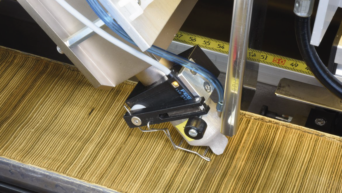
The lab is equipped with an X-Ray fluorescence core scanner to analyze split core sections up to 180 cm length and diameters between 6 and 12 cm. A CCD line-scan camera is used for optical line-scanning of the spit core samples with separate RGB sensors and UV-luminescence scanning of carbonate samples (e.g. speleothem, coral). Further, a micro X-Ray fluorescence scanner is used to measure polished and epoxy-embedded sediment samples, thick-sections and speleothem samples (sample stage 20 x 17 cm). Additionally, a Desktop-SEM with EDS can be used for uncoated samples upto 100 mm length.
Instruments
-
Micro-X-Ray Fluorescence Spectrometer
Measurement method of X-Ray fluorescence used to measure amounts of elements in a material. Micro-XRF (μ-XRF) analysis uses highly brilliant X-Ray sources (synchrotron source and spot size 100 nm to 2 μm) and microfocussing X-Ray optics to give fg to ag detection limits. (Source: IUPAC; https://doi.org/10.1515/pac-2019-0302)
-
Scanning Electron Microscope
The Scanning Electron Microscope (SEM) is one of the most versatile and widely used tools of modern science as it allows the study of both morphology and composition of biological and physical materials. By scanning an electron probe across a specimen, high resolution images of the morphology or topography of a specimen, with great depth of field, at very low or very high magnifications can be obtained. Compositional analysis of a material may also be obtained by monitoring secondary X-rays produced by the electron-specimen interaction. Thus detailed maps of elemental distribution can be produced from multi-phase materials or complex, bio-active materials. Characterization of fine particulate matter in terms of size, shape, and distribution as well as statistical analyses of these parameters, may be performed. There are many different types of SEM designed for specific purposes ranging from routine morphological studies, to high-speed compositional analyses or to the study of environment-sensitive materials. The Centre for Microscopy & Microanalysis presents three particular types of SEM that, in combination, provide a powerful analytical approach for many research or quality-control applications. Additional information available at "http://www.uq.edu.au/nanoworld/sem_gen.html" [Summary provided by The University of Queensland] (Source: Global Change Master Directory (GCMD). 2023. GCMD Keywords, Version 16.3. Greenbelt, MD: Earth Science Data and Information System, Earth Science Projects pision, Goddard Space Flight Center (GSFC) National Aeronautics and Space Administration (NASA). URL (GCMD Keyword Forum Page): https://forum.earthdata.nasa.gov/app.php/tag/GCMD+Keywords)
-
X-Ray Fluorescence Core Scanner